

ART Gallery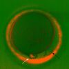
An oocyte extruding polar body (white arrow) possessing pseudo polar body (black arrow). Anaphase meiotic spindle visualized with polarized light microscopy. 21 December 2016 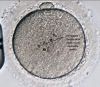
Refractile bodies are one of the main morphological abnormalities that can be observed in the cytoplasm of human oocytes. 20 December 2016 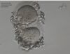
A Conjoined Oocytes Within One Follicle Have Infrequently Been Practical In Human Assisted Reproduction Thus Indication For The Hypothesis That Some Of The Polyovular Follicles Recognized From Histological Studies Of The Ovary May Persist Awaiting Ovulation. Image Illustrate Immature Oocytes In W 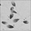
DNA Damage in Sperm Toroids revealed by Triton X-100 and DTT Pre-treatment before Staining. Sperm DNA compressed into toroids are exposed by acidified Triton X-100 and DTT pre-treatment (Biomed Res Int. 2015;2015:780983). DNA damage (darker color) is revealed following simple staining (Diff-Quik or Hema 3). "ROC calculated cut-off was >70.0% for normal toroid integrity (sensitiv 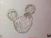
Implanted mouse embryo showing inner cell mass and trophoblasts. Implanted mouse embryo (Arch Gynecol Obstet. 2015;291:647) showing a dark central group of cells (inner cell mass or ICM) that becomes the fetus while the surrounding trophoblasts give rise to the placenta. 02 March 2016 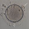
An egg with cellular fragment in Zona Pellucida An egg (outlined arrow indicates 1st polar body) with cell fragment (detached additional polar body??) embedded in Zona Pellucida (indicated by filled arrow) 29 February 2016 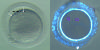
Immature oocyte with two germinal vesicles Left - stereomicroscope 40x, arrows indicate germinal vesicles. Right - same oocyte fixed and labelled for actin (phalloidin; cyan to white) and DNA (DAPI; maganta), confocal microscope 60x 29 February 2016 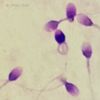
Simple Diff-Quik stain for chromatin integrity including DNA fragmentation. Dual use of Diff-Quik (Hema 3) for both strict morphology and chromatin integrity (Hum Reprod. 2009;24:28) can be improved (J Reprod Med. 2015;60:6) by an immediate post-stain 1-minute soak-in-water step to distinguish DNA damaged sperm which then appear as dark purple-blue sperm. This is due to 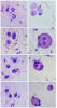
Difference between WBC and Spermatids 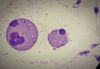
The multinuclear cell is a result of incomplete spermatogenesis resulting in undivided spermatids as an abnormal round cell. Spermatids with cytoplasm are at the stage just before sperm formation. Diff-Quik stained. 500x 16 February 2016 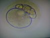
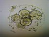
Zygote with multiple polar bodies PCOD patient over maturation resulted into polar body anomality but fertilized with IVF 13 July 2015 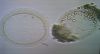
Day-3 Embryo With Thin Zona Pellucida- 4.5 microns A pregnancy by intracytoplasmic sperm injection (ICSI) of a couple whose oocytes were recovered without a zona pellucida is reported 17 July 2014 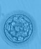
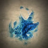
Fat Molecule-Found during Egg Pick Up-In Follicular Fluid Follicular fluid content(Fat Molecule) and oocyte quality13 July 2014 |