

IVF NewsNews: Protein that helps sperm fuse with egg identified
Dr George Janes 26 September 2022
A newly discovered protein which facilitates egg and sperm fusion provides new insights into human fertilisation. Over half of infertility cases cannot be explained, meaning that treatments based on current understanding may not be effective for every patient. Using an artificial fertilisation technique, researchers have revealed a novel protein controlling the attachment of sperm to eggs, shedding new light on the mechanisms of conception. Professor Harry Moore of the University of Sheffield and lead author of the study said: 'Infertility is unexplained in more than half of those who struggle to conceive naturally. What we know about fertility in humans has been severely limited by ethical concerns and the lack of eggs for research.' The study, published in Science Advances, describes how the team used microscopic beads, each coated with a specific protein fragment, to screen the role of many proteins in sperm-to-egg attachment. Using this method, they discovered that beads coated with a section of one protein, MAIA, named after the Greek goddess of motherhood, bound a high number of sperm. The scientists showed that MAIA acts together with a previously identified sperm-binding protein called JUNO to anchor sperm to the egg's surface. They introduced the genes for MAIA and JUNO into hamster egg cells and found that this enabled them to bind human sperm, something they ordinarily would not be able to do. PET trustee, Professor Allan Pacey of the University of Sheffield and co-author of the study said: 'This discovery of the MAIA protein is a major step forward in how we understand the process of human fertilisation. It would have been almost impossible to discover without the use of the artificial beads to replicate the surface of human eggs as we simply wouldn't have been able to get enough eggs to do the experiment. A classic case of thinking out of the box.' These findings elucidate how gamete fusion occurs and could explain the idea that, between humans, some people's sperm may not be compatible with others' eggs. The discovery of this mechanism and future work could help explain why some patients cannot easily conceive. Professor Moore commented: 'The ingenious artificial fertilisation technique which enabled us to identify the MAIA protein will not only allow scientists to better understand the mechanisms of human fertility but will pave the way for novel ways to treat infertility and revolutionise the design of future contraceptives.' Sources and References
[ Full Article ]  
News: Human cells mimicking early embryogenesis generated
Dr Emma Green 26 September 2022
A type of embryonic cell has been generated from human stem cells for the first time, providing a method to study post-implantation development. Published in Cell Stem Cell, researchers at KU Leuven, Belgium, have created extraembryonic mesoderm cells (EXMCs) from human induced pluripotent stem cells (iPSCs). The cells closely resemble those naturally formed in human embryos, providing a good model to study early development in vitro. 'We are very excited because now we can study processes that normally remain inaccessible during development,' said lead author Professor Vincent Pasque. 'The model has already enabled us to find out where extraembryonic mesoderm cells come from. In the longer term, our model will hopefully also shed more light on medical challenges such as fertility problems, miscarriages, and developmental disorders.' Ethical and technical limitations mean that early human embryo development is difficult to study. Human stem cell models provide an accessible way to study specific cells and their processes. Human iPSCs have the capacity to generate all cell types in the body. The extraembryonic mesoderm forms early in embryonic development just after implantation. EXMCs generate the first blood in the embryo, aid in attaching the embryo to the future placenta, and are involved in forming the primitive umbilical cord. 'In humans, this type of cell appears at an earlier developmental stage than in mouse embryos, and there might be other important differences between species. That makes our model especially important: research in mice may not give us answers that also apply to humans' said Professor Pasque. This new cell model is a step forward in understanding human cell development and may provide a way to study a variety of developmental disorders. Sources and References
[ Full Article ]
News: Hyaluronic acid may boost chance of live birth
Melinda Van Kerckvoorde 26 September 2022
A large data analysis study has found that embryo exposure to hyaluronic acid prior to transfer could improve IVF success rates for patients using their own eggs. Hyaluronic acid is an adhesive compound that is secreted by the cells surrounding the egg and is naturally present in the female reproductive tract. Following in-vitro fertilisation, embryos are kept in a liquid medium before being transferred back to the womb. Many studies have investigated whether adding hyaluronic acid to the culture medium could be a simple and cheap way to improve IVF success. A data analysis published in Human Reproduction suggests that it may increase live birth rates for some patients. 'We found that when women use their own eggs, exposing to hyaluronic acid for ten minutes before placing it in the uterus increased the likelihood of a cycle resulting in a birth by 32 percent to 39 percent,' said Dr Devorah Heymann from Kaplan Hospital in Rehovot, Israel who led the study in partnership with Hebrew University, Jerusalem, Israel. The research team analysed data from 15 clinical trials to compare the effect of hyaluronic acid on pregnancy outcomes between patients who used their own eggs or donor eggs for IVF. A total of 4686 IVF patients were included in the study whose embryo culture medium contained high or low concentrations or no hyaluronic acid. Following statistical analysis, the researchers claim that culture media containing high concentrations of hyaluronic acid increase the number of live births and clinical pregnancies from 36 percent to 43 percent and 42 to 47 percent, respectively, when using the patient's own eggs. No beneficial effect on pregnancy outcomes was observed when using donor eggs. The team had previously carried out a Cochrane Review in 2020 which had also shown that hyaluronic acid treatment could help people who use their own eggs for IVF. Given the increased demand for donor eggs, hyaluronic acid treatment thus could help patients to conceive even when their own eggs are of poorer quality. Furthermore, this study could provide more evidence for the Human Fertilisation and Embryology Authority which currently lists hyaluronic acid addition during IVF as a treatment add-on with conflicting results. The researchers highlight that more data is needed to reveal the true effect of hyaluronic acid when using donated eggs and that further research is required to understand the underlying mechanism of hyaluronic acid. Sources and References
[ Full Article ]
Announcement: IMT Matcher - IVF Witnessing System
EART 14 September 2022
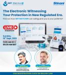
𝗦𝗵𝗶𝘃𝗮𝗻𝗶 𝗦𝗰𝗶𝗲𝗻𝘁𝗶𝗳𝗶𝗰 𝗶𝗻𝘃𝗶𝘁𝗲𝘀 𝗬𝗢𝗨 𝘁𝗼 𝗷𝗼𝗶𝗻 𝘂𝘀 𝗟𝗜𝗩𝗘 𝗼𝗻 𝟭𝟴𝘁𝗵 𝗦𝗲𝗽𝘁𝗲𝗺𝗯𝗲𝗿 𝟮𝟬𝟮𝟮, 𝗦𝗨𝗡𝗗𝗔𝗬 𝗮𝘁 𝟭𝟭:𝟬𝟬𝗮𝗺 [ Full Article ]
News: Further stages of development observed in mouse embryo models
Dr Rachel Montgomery 05 September 2022
'Synthetic' model embryos made using mouse stem cells and an artificial incubator are the closest a stem-cell-derived embryo model has ever come to resembling a naturally developing embryo in the uterus. Researchers from the University of Cambridge and the California Institute of Technology grew the so-called 'integrated stem cell-based embryo models' for 8.5 days (complete mouse gestation is 18-21 days) – long enough to develop a beating heart and the foundations of all other organs. 'The stem cell embryo model is important because it gives us accessibility to the developing structure at a stage that is normally hidden from us due to the implantation of the tiny embryo into the mother's womb' said lead researcher Professor Magdalena Zernicka-Goetz from the University of Cambridge. As reported in Nature, the models were developed by combining three different and specific types of stem cells: one that will develop into the embryo, with the other two developing into extraembryonic structures including the placenta and yolk sac which provides the early embryo with nutrients. When combined in an artificial incubator, the stem cells were found to signal to each other via touch and chemical messengers, allowing them to spontaneously self-organise into embryos. These synthetic models also reached the point where the entire brain, including the anterior forebrain, began to develop. 'The embryos struggle to grow beyond that point because they require a placenta which we cannot reproduce in vitro', first author Dr Gianluca Amadei from the University of Cambridge told the Financial Times. The recent work builds on very similar work published earlier this month by Professor Jacob Hanna at the Weizmann Institute of Science in Israel, who genetically altered stem cells to create mouse embryo models and also allowed them to develop for 8.5 days. Both studies found the process highly error-prone, with only one in 100 attempts being successful. Nonetheless, the team hope that their findings could have a number of implications, from improving the understanding of normal development and helping to better understand why some embryos go on to develop into a healthy pregnancy, while many others fail. The team are also working on developing human embryo models, but stress that these efforts are considerably further behind given the differences in understanding between early human and mouse development. Having a lab-grown human embryo model available for research could be a major advance for the study of fertility and common developmental disorders, but also highlight the importance of ongoing ethical and legal conversations about the status and use of model embryos and natural human embryos in research. Sarah Norcross, director of PET, said: 'Unlike actual human embryos, stem-cell-based embryo models in humans and other organisms are not restricted by a 14-day rule…These models are extremely helpful for research, but there still remains a case for changing the law to extend the 14-day rule, so that we can continue to learn from the precious embryos that have been kindly donated by fertility patients following their treatment.' Sources and References
[ Full Article ]
News: Frozen embryo IVF may increase childhood cancer risk
Michael Limmena 05 September 2022
Children born from IVF using frozen embryos may have a higher risk of developing childhood cancer according to a new study; however, the overall risk remains low. Frozen embryo transfer, sometimes called frozen-thawed embryo transfer, is a process in which embryos are frozen before being thawed and implanted for pregnancy. Worldwide, this type of fertility treatment is becoming increasingly more common; and children born from IVF using frozen embryos now exceeds those born using fresh embryos in many countries. 'A higher risk of cancer in children born after frozen-thawed embryo transfer in assisted reproduction, a large study from the Nordic countries found,' said co-author Professor Ulla-Britt Wennerholm from the University of Gothenburg, Sweden. [However] no increase in cancer was found among children born after assisted reproduction techniques overall,' she added. While elective freeze-all embryo cycles are becoming more common, the long-term medical risks of children being born from frozen embryos remain not well understood. In particular, there are many conflicting studies on the link between children being born using frozen embryos and a higher risk of developing childhood cancer. Scientists from the University of Gothenburg performed a cohort study using data from almost eight million Scandinavian children, publishing their findings in PLOS Medicine. The research team analysed the medical records from 7,944,248 million children born in Denmark, Norway, Sweden, and Finland between 1984 and 2015. Of these, 171,744 were born after IVF while the remaining 7,772,474 were not. Among those born after IVF, 22,630 were born using frozen-thawed embryos. The researchers found that these children were almost two times more likely to develop childhood leukaemia than those born using fresh embryos or natural conception. However, when frozen and fresh embryos were analysed as a single group, children born after IVF did not lead to a higher risk of childhood cancer. 'The large investigation of almost eight million Nordic children is highly impressive.' said CARE Fertility Group's chief scientific officer Dr Alison Campbell, who was not involved in the study. Despite the study's findings, the researchers emphasised that the results should be interpreted cautiously. Although the study is very large, it is important to note that out of the 22,630 children born after frozen-embryo transfer, only 48 of them later developed cancer. This limits the statistical strength of the study. Moreover, this study cannot determine causation and that the slightly elevated risk, according to the research team, may be due to many different factors wholly unrelated to frozen embryos. 'People who have children born following frozen embryo transfer should not be unduly concerned by the findings because the actual number of children affected by cancer, following frozen embryo transfer, is too small to draw firm conclusions,' noted Dr Campbell. Sources and References
[ Full Article ]
News: ART & Embryology training program
Chennai Fertility Center and Research Institute 03 September 2022
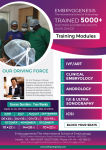
October 2022 Training Batch Schedule - 10th Oct - 22nd Oct 2022 The International School of Embryology was established to offer training for Clinicians in advanced Reproductive Technologies. Our skill and precision to all aspirants help them to know in-depth knowledge and experience. The members of our teaching faculty aim to bring doctors and embryologists to the highest level of knowledge about reproductive techniques and practical capability in the field. Our courses cover basics in Andrology, embryology, ICSI, and cryosciences (Hands-on). Limited Seats. For admission Contact 9003111598 / 8428278218 [ Full Article ]
News: Light shed on epigenetic maintenance of ovarian reserve
Melinda Van Kerckvoorde 22 August 2022
Egg cells are kept in a form of stasis from when they are first formed in the fetus until they mature in adulthood, by a protein which regulates transcription, a new study in mice has shown. During fetal development the ovarian reserve is formed which contains the early egg cells in the follicles. These cells are put into an arrested state immediately after entering the first phase of meiosis, a type of cell division which produces gametes, and may stay in this state for decades. How this reserve is established and maintained until the eggs 'ripen' has been poorly understood. 'Fertility is supported by these arrested oocytes,' said Professor Satoshi Namekawa, who led the study published in Nature Communications. 'The main question is how can these cells be maintained for decades? It's a big question. They cannot divide, they cannot proliferate, they just stay quiescent in the ovaries for decades. How is this possible?' The aim of this study was to find out if a dedicated epigenetic mechanism, which regulates which genes can be transcribed, regulates the formation and maintenance of the ovarian reserve in this meiotic state. Researchers looked at the potential role of a polycomb protein called PRC1, which is known to mediate epigenetic control of the genome. They specifically looked at what happened when the activity of that protein was silenced. When the PCR1 was silenced in genetically modified mice, ovaries were much smaller and contained fewer follicles, which is where the egg cells grow and mature. 'We show that a conditional PRC1 deletion results in rapid depletion of follicles and sterility, said Professor Namekawa. 'These results strongly implicate PRC1 in the critical process of maintaining the epigenome of primordial follicles throughout the protracted arrest that can last up to 50 years in humans'. Other findings demonstrated that PRC1 modulates the expression of many other genes during the formation of the ovarian reserve in the fetus, for example those involved in DNA repair and metabolic processes. Professor Namekawa concluded, 'Now that we found that this epigenetic process is key for establishment, the next question is can we uncover a more detailed mechanism of this process?' How can the ovarian reserve be maintained for decades?'. Finally, the research team hopes that future work will reveal if PRC1 dysfunction during this critical developmental window could explain premature ovarian failure and why human fertility declines with age. Sources and References
[ Full Article ]
News: Mitochondrial donation embryos appear to develop normally
Hannah Flynn 22 August 2022
Embryos produced using a method designed to allow women who are carriers of a mitochondrial condition to have genetic children without passing on their mitochondria, are similar to those produced using IVF with ICSI. Cells from embryos created using a form of mitochondrial donation called maternal spindle transfer had comparable levels of aneuploidy and genetic expression to control embryos created using ICSI, a recent study published in PLOS Biology by researchers from Peking University, Beijing, China showed. Professor Dusko Ilic, professor of stem cell science at King's College London, who was not involved in the research said: 'There is nothing surprising here. This study only further confirms that the spindle transfer is a safe technique for preventing transfer of mitochondrial mutations from mother to the embryo/child. The method has already been used in the clinic.' Maternal spindle transfer is just one of the methods that can be used to avoid mitochondrial DNA in a mother's egg from being transmitted to the subsequent embryo. In this method, nuclear material from the mother's egg is transferred to an unfertilised donor egg, which is then fertilised before the embryo is transferred to the uterus. Other methods exist which involve moving nuclear material shortly after fertilisation, rather than shortly before. The first baby born using maternal spindle transfer during IVF was born to a mother who was a carrier of Leigh Syndrome in 2016. Australia is the latest country to have legislated for the use of mitochondrial donation, having done so in March this year. Cells from 24 embryos created using this method were studied and it was found that 22 percent were aneupolid, meaning they had the wrong number of chromosomes, compared to 17 percent of cells studied from the 22 control embryos, created using ICSI. This was 'comparable' rate of aneuploidy the authors said. Cells taken from the three different layers of the blastocyst were also found to have RNA expression that was comparable to the control blastocysts created using ICSI, suggesting that the transcription of DNA was similar and not affected. DNA methylation levels were also similar in cells taken from the epiblast and primitive endoderm of the blastocysts of the embryos created using maternal spindle transfer when compared to the controls, but higher DNA methylation was observed in the trophectoderm. Authors suggested this could be due to a delay in methylation in embryos created using maternal spindle transfer, but proposed that the embryos could catch up. The authors also called for further research to closely monitor the health of children born via maternal spindle transfer, to determine the safety of the technology. Sources and References
[ Full Article ]
Podcast: Protect IVF!
International IVF Initiative 13 August 2022
A special podcast about the overturning in the US of Roe v Wade and its global implications. A joint venture with the International IVF Initiative (I3) and Doctors for Fertility- a group of reproductive endocrinology and infertility doctors with a mission to educate and inform policy on reproductive rights and to advocate and take political action for continued access to fertility treatment and preservation. Almost half a century ago, the Roe v. Wade ruling was the basis for establishing a constitutional right to abortion. The recent decision in Dobbs v. Jackson Women’s Health Organization demonstrates the increasingly conservative direction of the court in the US and prompts questions about the implications for civil rights, embryo rights, and health policy. What does this mean for IVF? It has raised fears that it could have "far-reaching ramifications" on people looking to get pregnant and the clinics providing services to help them. Will embryos created, frozen, used in fertility treatment, PGT, or discarded have rights? Will "trigger laws" go into effect that recognizes an embryo as a person? This closed zoom meeting with open dialogue involving a select panel of experts will offer content to help all that are impacted by this ruling. With examples of restrictive policies in the past and a call for action for all stakeholders, this podcast should support everyone that needs help and will offer advice to organize individuals and communities to fight restrictive IVF legislation in vulnerable States. Participants: Serena H Chen MD Giles Palmer David Sable MD Davina (Rudnick) Fankhauser Dr. Vivienne Hall Salu Ribeiro Lucky Sekhon MD Lowell Ku MD Colleen Quinn Stephanie Gustin MD Declan Keane Jacques Cohen PhD Lea Wilcox Cynthia Hudson Thomas Elliott Mary Ann Szvetecz Brad Zavy see also: Bill relating to clinical laboratories A New Challenge for Fertility Patients? Modeling IVF Access Post Repeal of Roe v. Wade The Solemn Truth About Medical Oaths [ Full Article ]
|