IVF NewsNews: Frozen embryo IVF may increase childhood cancer risk
Michael Limmena 05 September 2022
Children born from IVF using frozen embryos may have a higher risk of developing childhood cancer according to a new study; however, the overall risk remains low. Frozen embryo transfer, sometimes called frozen-thawed embryo transfer, is a process in which embryos are frozen before being thawed and implanted for pregnancy. Worldwide, this type of fertility treatment is becoming increasingly more common; and children born from IVF using frozen embryos now exceeds those born using fresh embryos in many countries. 'A higher risk of cancer in children born after frozen-thawed embryo transfer in assisted reproduction, a large study from the Nordic countries found,' said co-author Professor Ulla-Britt Wennerholm from the University of Gothenburg, Sweden. [However] no increase in cancer was found among children born after assisted reproduction techniques overall,' she added. While elective freeze-all embryo cycles are becoming more common, the long-term medical risks of children being born from frozen embryos remain not well understood. In particular, there are many conflicting studies on the link between children being born using frozen embryos and a higher risk of developing childhood cancer. Scientists from the University of Gothenburg performed a cohort study using data from almost eight million Scandinavian children, publishing their findings in PLOS Medicine. The research team analysed the medical records from 7,944,248 million children born in Denmark, Norway, Sweden, and Finland between 1984 and 2015. Of these, 171,744 were born after IVF while the remaining 7,772,474 were not. Among those born after IVF, 22,630 were born using frozen-thawed embryos. The researchers found that these children were almost two times more likely to develop childhood leukaemia than those born using fresh embryos or natural conception. However, when frozen and fresh embryos were analysed as a single group, children born after IVF did not lead to a higher risk of childhood cancer. 'The large investigation of almost eight million Nordic children is highly impressive.' said CARE Fertility Group's chief scientific officer Dr Alison Campbell, who was not involved in the study. Despite the study's findings, the researchers emphasised that the results should be interpreted cautiously. Although the study is very large, it is important to note that out of the 22,630 children born after frozen-embryo transfer, only 48 of them later developed cancer. This limits the statistical strength of the study. Moreover, this study cannot determine causation and that the slightly elevated risk, according to the research team, may be due to many different factors wholly unrelated to frozen embryos. 'People who have children born following frozen embryo transfer should not be unduly concerned by the findings because the actual number of children affected by cancer, following frozen embryo transfer, is too small to draw firm conclusions,' noted Dr Campbell. Sources and References
[ Full Article ] News: ART & Embryology training program
Chennai Fertility Center and Research Institute 03 September 2022

October 2022 Training Batch Schedule - 10th Oct - 22nd Oct 2022 The International School of Embryology was established to offer training for Clinicians in advanced Reproductive Technologies. Our skill and precision to all aspirants help them to know in-depth knowledge and experience. The members of our teaching faculty aim to bring doctors and embryologists to the highest level of knowledge about reproductive techniques and practical capability in the field. Our courses cover basics in Andrology, embryology, ICSI, and cryosciences (Hands-on). Limited Seats. For admission Contact 9003111598 / 8428278218 [ Full Article ] News: Light shed on epigenetic maintenance of ovarian reserve
Melinda Van Kerckvoorde 22 August 2022
Egg cells are kept in a form of stasis from when they are first formed in the fetus until they mature in adulthood, by a protein which regulates transcription, a new study in mice has shown. During fetal development the ovarian reserve is formed which contains the early egg cells in the follicles. These cells are put into an arrested state immediately after entering the first phase of meiosis, a type of cell division which produces gametes, and may stay in this state for decades. How this reserve is established and maintained until the eggs 'ripen' has been poorly understood. 'Fertility is supported by these arrested oocytes,' said Professor Satoshi Namekawa, who led the study published in Nature Communications. 'The main question is how can these cells be maintained for decades? It's a big question. They cannot divide, they cannot proliferate, they just stay quiescent in the ovaries for decades. How is this possible?' The aim of this study was to find out if a dedicated epigenetic mechanism, which regulates which genes can be transcribed, regulates the formation and maintenance of the ovarian reserve in this meiotic state. Researchers looked at the potential role of a polycomb protein called PRC1, which is known to mediate epigenetic control of the genome. They specifically looked at what happened when the activity of that protein was silenced. When the PCR1 was silenced in genetically modified mice, ovaries were much smaller and contained fewer follicles, which is where the egg cells grow and mature. 'We show that a conditional PRC1 deletion results in rapid depletion of follicles and sterility, said Professor Namekawa. 'These results strongly implicate PRC1 in the critical process of maintaining the epigenome of primordial follicles throughout the protracted arrest that can last up to 50 years in humans'. Other findings demonstrated that PRC1 modulates the expression of many other genes during the formation of the ovarian reserve in the fetus, for example those involved in DNA repair and metabolic processes. Professor Namekawa concluded, 'Now that we found that this epigenetic process is key for establishment, the next question is can we uncover a more detailed mechanism of this process?' How can the ovarian reserve be maintained for decades?'. Finally, the research team hopes that future work will reveal if PRC1 dysfunction during this critical developmental window could explain premature ovarian failure and why human fertility declines with age. Sources and References
[ Full Article ] News: Mitochondrial donation embryos appear to develop normally
Hannah Flynn 22 August 2022
Embryos produced using a method designed to allow women who are carriers of a mitochondrial condition to have genetic children without passing on their mitochondria, are similar to those produced using IVF with ICSI. Cells from embryos created using a form of mitochondrial donation called maternal spindle transfer had comparable levels of aneuploidy and genetic expression to control embryos created using ICSI, a recent study published in PLOS Biology by researchers from Peking University, Beijing, China showed. Professor Dusko Ilic, professor of stem cell science at King's College London, who was not involved in the research said: 'There is nothing surprising here. This study only further confirms that the spindle transfer is a safe technique for preventing transfer of mitochondrial mutations from mother to the embryo/child. The method has already been used in the clinic.' Maternal spindle transfer is just one of the methods that can be used to avoid mitochondrial DNA in a mother's egg from being transmitted to the subsequent embryo. In this method, nuclear material from the mother's egg is transferred to an unfertilised donor egg, which is then fertilised before the embryo is transferred to the uterus. Other methods exist which involve moving nuclear material shortly after fertilisation, rather than shortly before. The first baby born using maternal spindle transfer during IVF was born to a mother who was a carrier of Leigh Syndrome in 2016. Australia is the latest country to have legislated for the use of mitochondrial donation, having done so in March this year. Cells from 24 embryos created using this method were studied and it was found that 22 percent were aneupolid, meaning they had the wrong number of chromosomes, compared to 17 percent of cells studied from the 22 control embryos, created using ICSI. This was 'comparable' rate of aneuploidy the authors said. Cells taken from the three different layers of the blastocyst were also found to have RNA expression that was comparable to the control blastocysts created using ICSI, suggesting that the transcription of DNA was similar and not affected. DNA methylation levels were also similar in cells taken from the epiblast and primitive endoderm of the blastocysts of the embryos created using maternal spindle transfer when compared to the controls, but higher DNA methylation was observed in the trophectoderm. Authors suggested this could be due to a delay in methylation in embryos created using maternal spindle transfer, but proposed that the embryos could catch up. The authors also called for further research to closely monitor the health of children born via maternal spindle transfer, to determine the safety of the technology. Sources and References
[ Full Article ] Podcast: Protect IVF!
International IVF Initiative 13 August 2022

A special podcast about the overturning in the US of Roe v Wade and its global implications. A joint venture with the International IVF Initiative (I3) and Doctors for Fertility- a group of reproductive endocrinology and infertility doctors with a mission to educate and inform policy on reproductive rights and to advocate and take political action for continued access to fertility treatment and preservation. Almost half a century ago, the Roe v. Wade ruling was the basis for establishing a constitutional right to abortion. The recent decision in Dobbs v. Jackson Women’s Health Organization demonstrates the increasingly conservative direction of the court in the US and prompts questions about the implications for civil rights, embryo rights, and health policy. What does this mean for IVF? It has raised fears that it could have "far-reaching ramifications" on people looking to get pregnant and the clinics providing services to help them. Will embryos created, frozen, used in fertility treatment, PGT, or discarded have rights? Will "trigger laws" go into effect that recognizes an embryo as a person? This closed zoom meeting with open dialogue involving a select panel of experts will offer content to help all that are impacted by this ruling. With examples of restrictive policies in the past and a call for action for all stakeholders, this podcast should support everyone that needs help and will offer advice to organize individuals and communities to fight restrictive IVF legislation in vulnerable States. Participants: Serena H Chen MD Giles Palmer David Sable MD Davina (Rudnick) Fankhauser Dr. Vivienne Hall Salu Ribeiro Lucky Sekhon MD Lowell Ku MD Colleen Quinn Stephanie Gustin MD Declan Keane Jacques Cohen PhD Lea Wilcox Cynthia Hudson Thomas Elliott Mary Ann Szvetecz Brad Zavy see also: Bill relating to clinical laboratories A New Challenge for Fertility Patients? Modeling IVF Access Post Repeal of Roe v. Wade The Solemn Truth About Medical Oaths [ Full Article ] News: Placenta and organ formation observed in mouse embryo models
Dr Rachel Montgomery 08 August 2022
Researchers in Israel have observed the formation of the placenta, yolk sac and some organs, in embryo models derived entirely from mouse embryonic stem cells. The research was performed at the Weizmann Institute of Science in Rehovot, and was published in the journal Cell. Previous work by the team published in Nature last year had focused on developing ways to successfully grow embryos outside of the uterus, or ex utero, a process employed again for this study. However previous work by the team had focused on growing embryos removed from a mouse uterus after two days, in the simulated uterus they developed, whereas their latest study shows embryo models made entirely from embryonic stem cells could reach the stage where organs start to form, outside of the uterus. They were also able to induce embryonic stem cells to produce cells and structures that would go on to develop into the placenta, amniotic membrane and yolk sac. Lead researcher Professor Jacob Hanna said: 'Until now, in most studies, the specialised cells were often either hard to produce or aberrant, and they tended to form a mishmash instead of well-structured tissue suitable for transplantation. We managed to overcome these hurdles by unleashing the self-organisation potential encoded in the stem cells.' To develop the structures, the researchers took mouse embryonic stem cells, and divided them into three groups. One group was destined to develop into embryonic organs, while the other two were treated in a way to give rise to either placental cells or the yolk sac – so-called extra-embryonic structures that are required to support the developing embryo. All three cell types were then mixed together, and incubated in the artificial womb the team had previously developed. Soon after being mixed together, the cells self-organised into aggregates. However, overall the experiment was highly error-prone as 99.5 percent of these aggregates failed to develop further. Nonetheless, 0.5 percent (around 50 of 10,000 aggregates) did go on to form spheres, which later changed further into elongated, embryo-like structures. These were allowed to develop for eight and a half days, a third of the gestation of a mouse. By this point, they had formed a beating heart, blood stem cell circulation, rudimentary brain with folds, a neural tube and a gut tube. Gene expression patterns in the embryo models were mapped and researchers found they were 95 percent similar to natural mouse models. Dr James Briscoe, principal group leader and assistant research director, at the Francis Crick Institute, said: 'The study has broad implications as although the prospect of synthetic human embryos is still distant, it will be crucial to engage in wider discussions about the legal and ethical implications of such research.' Sources and References
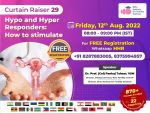
[ Full Article ] News: Topic - 𝙃𝙮𝙥𝙤 & 𝙃𝙮𝙥𝙚𝙧 𝙍𝙚𝙨𝙥𝙤𝙣𝙙𝙚𝙧𝙨: 𝙃𝙤𝙬 𝙩𝙤 𝙨𝙩𝙞𝙢𝙪𝙡𝙖𝙩𝙚?
Dr. Prof (Col) Pankaj Talwar VSM 08 August 2022

✅ Coming Friday 12th Aug 2022 - 𝙇𝙞𝙫𝙚 𝙯𝙤𝙤𝙢 𝙨𝙚𝙨𝙨𝙞𝙤𝙣 - Evening 7:45p𝙢 (𝙄𝙎𝙏)
𝙎𝙥𝙚𝙖𝙠𝙚𝙧 - 𝘿𝙧. 𝙋𝙧𝙤𝙛. (𝘾𝙤𝙡) 𝙋𝙖𝙣𝙠𝙖𝙟 𝙏𝙖𝙡𝙬𝙖𝙧, 𝙑𝙎𝙈!
Topic - 𝙃𝙮𝙥𝙤 & 𝙃𝙮𝙥𝙚𝙧 𝙍𝙚𝙨𝙥𝙤𝙣𝙙𝙚𝙧𝙨: 𝙃𝙤𝙬 𝙩𝙤 𝙨𝙩𝙞𝙢𝙪𝙡𝙖𝙩𝙚?
#𝙅𝙤𝙞𝙣 𝙎𝙥𝙡.𝙒𝙝𝙖𝙩𝙨𝙖𝙥𝙥 𝙂𝙧𝙤𝙪𝙥 𝙩𝙤 𝙜𝙚𝙩 𝙕𝙤𝙤𝙢 𝙎𝙚𝙨𝙨𝙞𝙤𝙣 𝙞𝙣𝙫𝙞𝙩𝙚 𝙡𝙞𝙣𝙠: https://chat.whatsapp.com/ClRzEOauuRfBAbru5kJQgx
𝙊𝙋𝙀𝙉 𝙁𝙊𝙍 𝘼𝙡𝙡!
𝙁𝙤𝙧 𝙁𝙧𝙚𝙚 𝙍𝙚𝙜𝙞𝙨𝙩𝙧𝙖𝙩𝙞𝙤𝙣 𝙤𝙧 𝙢𝙤𝙧𝙚 𝙞𝙣𝙛𝙤 :- https://wa.me/918287883005
#hyporesponders #ovulationinduction #iui #ivf #onlineivfcourses #elearning #webinar #MD #MS #IVFCourses #onlinecourses #ivfonlinecourses #reproductiveultrasound #usg #et #OPU #iceat #OBG #DGO #gynaecologist #obstetrics #certification #semenanalysis #ART #ARTTraining #EmbryologyTraining #Andrology [ Full Article ] News: ART & Embryology training program
Chennai Fertility Center and Research Institute 05 August 2022

September 2022 Training Batch Schedule - 5th Sep - 19th Sep 2022 The International School of Embryology was established to offer training for clinicians in advanced reproductive technologies. Our skill and precision to all aspirants help them to know in-depth knowledge and experience. The members of our teaching faculty aim to bring doctors and embryologists to the highest level of knowledge about reproductive techniques and practical capability in the field. Our courses cover basics in Andrology, embryology, ICSI, and cryosciences (Hands-on). Limited Seats. For admission Contact 9003111598 / 8428278218 [ Full Article ] News: Differences in IVF-conceived children's size disappear by adolescence
Dr Maria Botcharova 03 August 2022
A large multi-cohort study has shown that adults conceived following fertility treatment have no difference in height, weight, and body fat to those who were conceived naturally. While children conceived via IVF and ICSI were shorter, lighter and thinner than their naturally conceived counterparts, these differences were shown to disappear by the time both groups reached late adolescence. 'This is important work,' said Dr Ahmed Elhakeem, senior research associate in epidemiology at Bristol University and lead author of the paper. 'Parents and their children conceived by [assisted reproductive technologies] ART can be reassured that this might mean they are a little bit smaller and lighter from infancy to adolescence, but these differences are unlikely to have any health implications'. The study published in JAMA Network Open aligns with previous research, which showed children conceived via IVF have increased risk of preterm birth, as well as being smaller and lighter, but which have mostly focused on the perinatal period, in the first year of the child's life. It used data collated from 158,066 participants from Europe, Asia and Canada, aged between several weeks and 27 years. Of these 4329 had been conceived via IVF or ICSI. 'This important research is only possible through large scale international collaboration and longitudinal health studies, where participants contribute health data throughout their entire lives,' said Professor Deborah Lawlor, another author of the study, MRC Investigator and British Heart Foundation chair. The study showed that any differences in height, weight and BMI diminished steadily as children grew older, with the difference completely diminished in adolescents aged between 14 to 17 years. A similar pattern was seen in weight, waist circumference, body fat percentage, and fat mass index showed similar results to BMI. Since its first use in 1978 IVF has contributed to over eight million births globally. 'In the UK just over one in 30 children have been conceived by ART, so we would expect on average one child in each primary school class to have been conceived this way.' Dr Elhakeem commented. Peter Thompson, chief executive of the Human Fertilisation and Embryology Authority added 'Around one in seven couples have difficulty conceiving in the UK which leads to around 53,000 patients a year having fertility treatment (IVF or donor insemination). The findings from this study will come as a welcome relief to these patients who begin treatment in the hope of one day having healthy children of their own'. Sources and References
[ Full Article ] News: Egg cells remain dormant for decades by putting mitochondria to sleep
Eleanor Gallegos 03 August 2022
Early human oocytes remodel their metabolic activity, which enables them to remain dormant and reproductively viable for decades. Humans form oocytes during fetal development. These oocytes then undergo cellular arrest and remain dormant in the ovaries for up to 50 years. During dormancy the oocytes maintain mitochondrial activity to generate energy for essential cell processes. This energy generation produces reactive oxygen species (ROS) as by-products. ROS are highly reactive oxygen-containing molecules which are harmful in high concentrations and can damage oocytes causing cell death. A paper published in Nature has revealed that oocytes can alter their metabolic pathways to limit the production of ROS. 'Humans are born with all the supply of egg cells they have in life. As humans are also the longest-lived terrestrial mammal, egg cells have to maintain pristine conditions while avoiding decades of wear-and-tear.' said Dr Aida Rodriguez, postdoctoral researcher at the Centre for Genomic Regulation (CRG) in Barcelona, Spain, and first author of the study. 'We show this problem is solved by skipping a fundamental metabolic reaction that is also the main source of damage to the cell. As a long-term maintenance strategy, it's like putting batteries on standby mode. This represents a brand new paradigm never before seen in animal cells,'. The researchers studied Xenopus (African clawed frog) and human oocytes in early- and late-stages of development. Live imaging showed that early human and Xenopus oocytes do not generate any detectable ROS signal. Mitochondria contain five complexes, I-V, which perform the chemical reactions needed to generate energy in a cell. The researchers inhibited each of these complexes in Xenopus oocytes and found that while early- and late-stage oocytes died upon inhibition of complexes II-V, 78 percent of early-stage oocytes survived if complex I was inhibited, suggesting this complex is not used at this stage of development. The researchers then examined whether the subunits which make up complex I are depleted in early human oocytes and found that they were either absent or at very low levels. When studying ROS levels and complex I assembly, they found that ROS start to build up as complex I is formed. These results show that complex I is absent in early human oocytes which limits ROS production and the related cell damage. This metabolic remodelling helps maintain the reproductive capacity of early oocytes for the years in which they remain dormant. The researchers have proposed that the absence of complex I in early human oocytes could be exploited for other purposes such as cancer treatment. 'Complex I inhibitors have previously been proposed as a cancer treatment. If these inhibitors show promise in future studies, they could potentially target cancerous cells while sparing oocytes,' explained senior author Dr Elvan Böke, group leader in the Cell and Developmental Biology programme at the CRG. These results could have life-changing effects on the quality of life of young women post cancer treatment by avoiding infertility which can result from chemotherapy. Sources and References
[ Full Article ] |